44 ear anatomy without labels
› en › e-AnatomyNormal chest MDCT with anatomic labels | e-Anatomy - IMAIOS Mar 10, 2022 · Normal anatomy of the thorax on labeled Chest CT: radiological anatomy of the lungs, mediastinal lymph nodes, trachea, bronchi, pleural cavity, heart and pulmonary vessels. × Your email address is not verified. Bones Of The Human Body Without Labels at Anatomy Bones Of The Human Body Without Labels. They range in size from the tiniest found in the middle ear, to the largest that forms our thigh.the human body has an amazing array of different bones, many of which you can find on yourself or on a skeleton.knowledge of the skeletal structure of the human body is essential to know before.
Skeletal System Diagram Without Labels - Anatomy Organ Apr 5, 2018 - Skeletal System Diagram Without Labels - See more about Skeletal System Diagram Without Labels, skeletal system diagram no labels, skeletal system diagram without labels, skeletal system picture without label

Ear anatomy without labels
The Ear: Anatomy, Function, and Treatment - Verywell Health Essential organs of human hearing and balance, the ears are located on either side of the head, at the level of the nose. Separated into an inner, middle, and outer ear, each ear is an intricate and complicated mixture of bones, nerves, and muscles. Anatomy of the Ear | Geeky Medics The tympanic membrane, or eardrum, marks the border between the external and middle ear. It is formed of a middle layer of connective tissue with a layer of skin on its lateral surface (facing the external acoustic meatus) and mucous membrane on its medial surface (facing the middle ear). en.wikipedia.org › wiki › Human_penisHuman penis - Wikipedia The distal section of the urethra allows a human male to direct the stream of urine by holding the penis. This flexibility allows the male to choose the posture in which to urinate. In cultures where more than a minimum of clothing is worn, the penis allows the male to urinate while standing without removing much of the clothing.
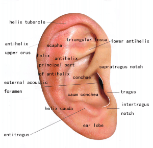
Ear anatomy without labels. Blank ear diagrams and quizzes: The fastest way to learn ... Ear diagrams (labeled and unlabeled) Accelerate your learning with interactive quizzes Sources + Show all Ear anatomy overview Although it's not obvious to look at, the ear is anatomically divided into three portions: External (outer) ear Middle ear Inner ear As you can imagine, there's a lot of associated anatomy to learn for each portion! Outer Ear: Anatomy, Location, and Function - Verywell Health Helix: The outermost curvature of the ear, extending from where the ear joins the head at the top to where it meets the lobule. The helix begins the funneling of sound waves into the ear; Fossa, superior crus, inferior crus, and antihelix: These sections make up the middle ridges and depressions of the outer ear. The superior crus is the first ridge that emerges moving in from the helix. Ear Diagram and Labeling Worksheet / Worksheet The first worksheet presents an ear with annotations showing the first letters of its key features. For example, a label marked 'P' links to the Pinna (outer ear). The second page shows an ear diagram without labels. The final page shows the labels linking to the beginning letters of each feature, but without the words list. Label Anatomy Teaching Resources | Teachers Pay Teachers Anatomy Lab Cabinet Labels Room Decor - Script font. by. Teaching is a Lifestyle with Seeds of Study. $1.00. PDF. Human anatomy cabinet labels for anatomy models. Script font.Print on cardstock, cut out and stick to your cabinets. Each label is 4cmx18cmThe labels come with (and without) the word "system."
› en › e-Anatomye-Anatomy: radiologic anatomy atlas of the human body - IMAIOS e-Anatomy is an award-winning interactive atlas of human anatomy. It is the most complete reference of human anatomy available on web, iPad, iPhone and android devices. Explore over 6700 anatomic structures and more than 870 000 translated medical labels. Images in: CT, MRI, Radiographs, Anatomic diagrams and nuclear images. Available in 12 ... Parts of the Ear Labelled Diagram Activity - Twinkl The first worksheet presents an ear with annotations showing the first letters of its key features. For example, a label marked 'P' links to the Pinna (outer ear). The second page shows an ear diagram without labels. The final page shows the labels linking to the beginning letters of each feature, but without the words list. Human Body Parts Images Without Labels - Free Vector ... Human ear diagram with labels and label of anatomy labeling the ear purposegames nose diagram with label diagrams all labels human ear the ear diagram without labels anatomy human charts. Illustration Of Body Parts Labels It is certainly the most widely studied structure the world over. Human body parts images without labels. Download body ... en.wikipedia.org › wiki › Anatomical_terminologyAnatomical terminology - Wikipedia Anatomy is often described in planes, referring to two-dimensional sections of the body. A section is a two-dimensional surface of a three-dimensional structure that has been cut. A plane is an imaginary two-dimensional surface that passes through the body. Three planes are commonly referred to in anatomy and medicine:: 4
Ear Anatomy - Outer Ear | McGovern Medical School The medical term for the outer ear is the auricle or pinna. The outer ear is made up of cartilage and skin. There are three different parts to the outer ear; the tragus, helix and the lobule. EAR CANAL The ear canal starts at the outer ear and ends at the ear drum. The canal is approximately an inch in length. Image result for ear structure without label | Ear diagram ... Feb 12, 2018 - Image result for ear structure without label. Feb 12, 2018 - Image result for ear structure without label. Pinterest. Today. Explore. ... Label Ear Anatomy Diagram Printout. G. Kim Gregory. Science. Human Brain Anatomy. Anatomy And Physiology. Animal Coloring Pages. Coloring Book Pages. Picture of the Ear: Ear Conditions and Treatments - WebMD WebMD's Ear Anatomy Page provides a detailed image and definition of the ear as well as an overview of ear-related health problems. Learn about the ear's function in the body and test and ... The Human Ear - Structure, Functions and its Parts - BYJUS For more information on the human ear, the structure of ear class 9, human ear parts, the function of the ear, and other related topics keep visiting BYJU'S website or download BYJU'S app for further reference. Learn more in detail about human ear, the structure, functions and other related topics at BYJU'S Biology.

picture front of the eye without labels clipart - Clipground
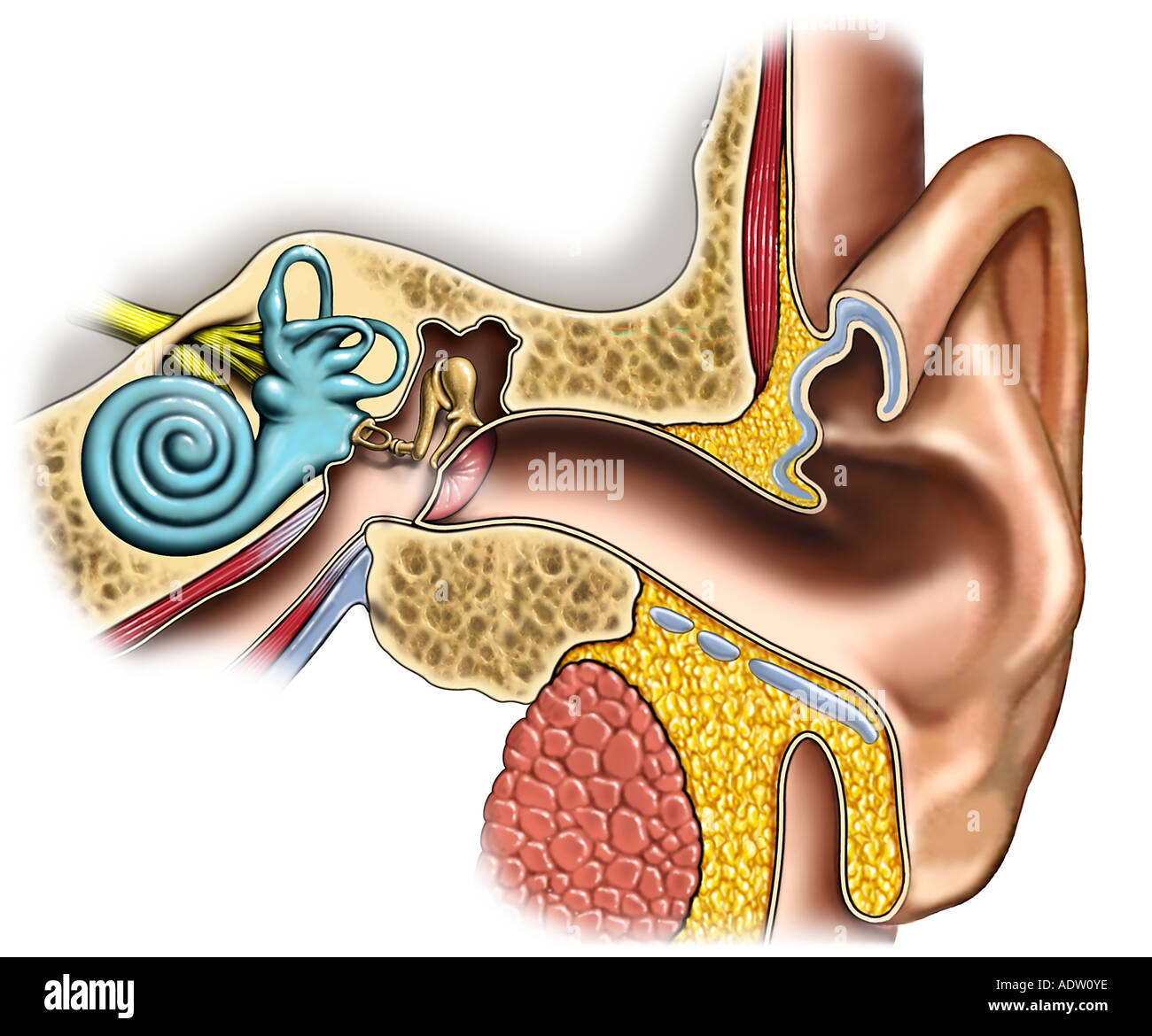
Anatomy and Physiology of the Ear - University of Rochester Pinna or auricle. This is the outside part of the ear. External auditory canal or tube. This is the tube that connects the outer ear to the inside or middle ear. Tympanic membrane (eardrum). The tympanic membrane divides the external ear from the middle ear. Middle ear (tympanic cavity), consisting of: Ossicles.
Exam Macros - TRAUMA YELLOW
Ear Anatomy: Understanding the Outer, Middle, and Inner ... The external auditory meatus, or ear canal, is a narrow canal that leads from the concha to the tympanic membrane, or eardrum. Sound waves are delivered through this canal. This canal is prone to ear infections. Tragus This is the small, rigid part of the ears along the front of the ear, adjacent to the face.
Ears - Pasadena & Newport Beach | Dr. Hung
Human Ear Anatomy - Parts of Ear Structure, Diagram and ... The external (outer) ear consists of the auricle, external auditory canal, and eardrum (Figure 1 and 2). The auricle or pinna is a flap of elastic cartilage shaped like the flared end of a trumpet and covered by skin. The rim of the auricle is the helix; the inferior portion is the lobule. Ligaments and muscles attach the auricle to the head.

How Do We Hear? | Maine Academy of Audiology
"Outer Ear Anatomy Colorful Pattern Without Center Label" Shop "outer ear anatomy colorful pattern without center label" search results for the very best in custom shoes, sneakers, apparel, and accessories by independent artists.

Human Ear Diagram with Label - coordstudenti
Human Eye Diagram Without Labels - solved label this ... Human Eye Diagram Without Labels - 13 images - picture front of the eye without labels clipart clipground, diagram of the eye clipart etc, eye diagram images stock photos vectors shutterstock, eye anatomy images stock photos vectors shutterstock,

Frequently Asked Questions - VeDA
Well-Labelled Diagram Of Ear With Explanation - BYJUS Eustachian Tube is a tube that connects the middle ear to the back of the nose. It helps to maintain equal pressure in the middle ear which facilitates the proper transmission of sound waves. The Inner ear consists of: Cochlea that comprises the nerves of hearing. Semicircular canals that contain the receptors that help in maintaining balance.
귀 해부 Ear Anatomy
Anatomy of the Ear | Inner Ear | Middle Ear | Outer Ear Anatomy of the Ear. The ear is made up of three parts: the outer, middle, and inner ear. All three parts of the ear are important for detecting sound by working together to move sound from the outer part through the middle and into the inner part of the ear. Ears also help to maintain balance.
Anatomy of the Left Ear: Cross-Section Stock Photo, Royalty Free Image: 7711165 - Alamy
Label Parts of the Human Ear - University of Dayton Label Parts of the Human Ear. Select One Auditory Canal Cochlea Cochlear Nerve Eustachian Tube Incus Malleus Oval Window Pinna Round Window Semicircular Canals Stapes Tympanic Membrane Vestibular Nerve. Select One Auditory Canal Cochlea Cochlear Nerve Eustachian Tube Incus Malleus Oval Window Pinna Round Window Semicircular Canals Stapes ...

Anatomy of the ear | Sketchy Medicine
Ear Anatomy Without Labels Digital Art Stock Illustration ... Ear Anatomy Without Labels Digital Art Stock Illustration 530108302 Edit Download for free See more Popularity score High Usage score High usage Superstar Shutterstock customers love this asset! Item ID: 530108302 Ear Anatomy without Labels, Digital Art Formats 8976 × 6201 pixels • 29.9 × 20.7 in • DPI 300 • JPG

Audiology & Speech Pathology 303 > Hedrick > Flashcards > Exam 1 AUSP 303 | StudyBlue
Human Ear Diagram - Bodytomy Auditory Ossicles: The three small bones in the middle ear, called malleus, stapes, and incus, are connected. These bones together are called the auditory ossicles, and their purpose is to let the sound that strikes the eardrum, further into the inner ear.

72 best N U R S I N G images on Pinterest | Nursing schools, Schools for nursing and Student nurse
en.wikipedia.org › wiki › Human_penisHuman penis - Wikipedia The distal section of the urethra allows a human male to direct the stream of urine by holding the penis. This flexibility allows the male to choose the posture in which to urinate. In cultures where more than a minimum of clothing is worn, the penis allows the male to urinate while standing without removing much of the clothing.

Hearing Health – Impact_Hearing
Anatomy of the Ear | Geeky Medics The tympanic membrane, or eardrum, marks the border between the external and middle ear. It is formed of a middle layer of connective tissue with a layer of skin on its lateral surface (facing the external acoustic meatus) and mucous membrane on its medial surface (facing the middle ear).

Unlabeled Digestive System Diagram Without Labels ~ news word
The Ear: Anatomy, Function, and Treatment - Verywell Health Essential organs of human hearing and balance, the ears are located on either side of the head, at the level of the nose. Separated into an inner, middle, and outer ear, each ear is an intricate and complicated mixture of bones, nerves, and muscles.
Anatomy of the Pharynx Medical Illustrations — Campbell Medical Illustration
ear

Ear | Diseases

Structure and Functions of the Ear Explicated With Diagrams
Post a Comment for "44 ear anatomy without labels"